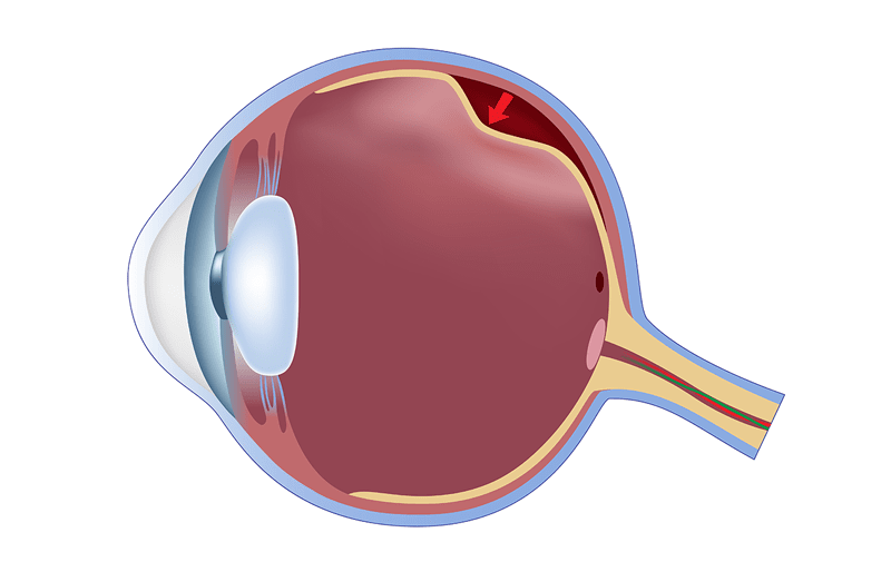
At retinal level complicated processes allow us to record images.
Central area (macula) records images in bright light (cones) giving us the possibility to detect fine details. Signals from the cones are sent to the brain where these messages are translated into the perception of colors.
Middle and peripheral retina is useful for vision in the presence of low light (rods).
Rods are used to detect change between light and dark and to recognize shape and movement detection.
The retina must have anatomical and functional connections with the surrounding tissues to work properly.
The retina is a part of the nervous system and can’t regenerate.
If the retina remains detached for a long time retinal cells die and the functional recovery is poor.
There are 3 possible types of retinal detachment (tractional, regmatogenous and exudative).
It is necessary to diagnose and treat retinal detachment in the shortest time possible.
If a retinal detachment has occurred the only possible therapy is surgical (retinopexy, cerclage with or without plumbing, vitrectomy).
The diagnosis of retinal detachment is done with eye examination and with ocular fundus examination in mydriasis ( pupil dilatation).


