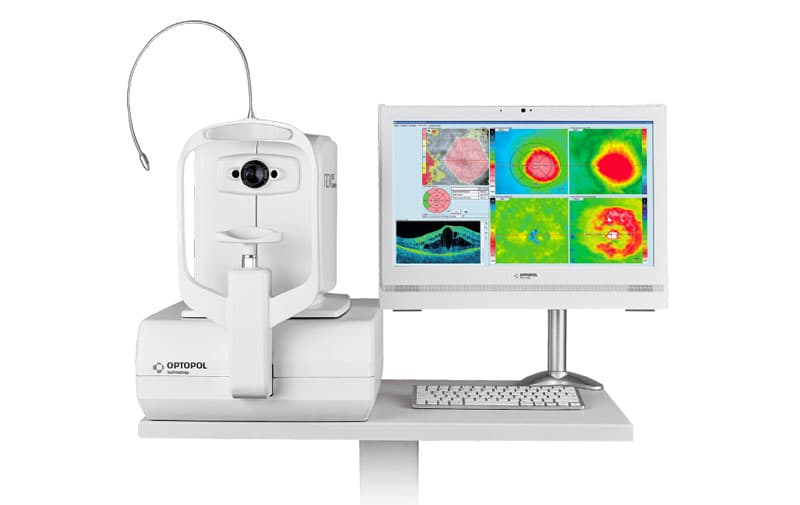
It is used to obtain images of the cornea, of the iridosclerocorneal angle, of the macular region, of the head of the optic nerve.
This is a non-invasive, fast-performing exam that gives the doctor a lot of useful information on the anatomy of the eye and can improve diagnosis, therapy and follow-up.
No harmful radiation is used.
It is not necessary to instill drops.
The examination lasts about 15 minutes.
May need this exam:
- Patients with suspected glaucoma or not able to perform visual field exam
- Diabetic patients with possible macular edema
- Patients with previous retinal thrombosis to evaluate presence and degree of a macular edema
- Patients with maculopathy to evaluate retinal involvment and assess presence of any complications
- Patients with macular pucker to evaluate retinal thickness and presence of alterations in the retina structure
- Patients with macular hole to evaluate and measure the size of the hole
- Myopic patients to evaluate presence of a myopic maculopathy
- Patients with a central serous chorioretinitis
- Patients who have a visual loss and an inconsistent objectivity
- We use the OCT REVO Copernicus for the examination


